
Ayurveda
Abhyanga Snanam – Ayurveda’s take
For many out there Diwali is the festival of lights. But If you ask me what is Diwali, I would say “Diwali is a wisely
As a mother, I always had a hundred thoughts in my mind during my pregnancy. I always wanted to be sure about whether my baby is growing well, whether she has any defects in the organs, are there any tests to assure myself about her wellbeing. This scan was a big relief then.
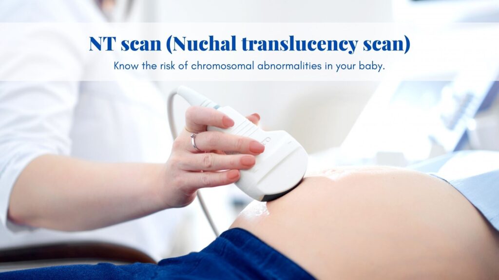
Yes! The results of an NT Scan (nuchal translucency scan) tell you if your baby has a high or low risk of a chromosomal abnormality if any.
Now you may wonder why this NT is measured. So increased thickness of the nuchal translucency might indicate a chromosomal abnormality, but it doesn’t tell you that your baby definitely has, or doesn’t have, an abnormality. The results will tell you if your baby is at high risk or low risk of chromosomal abnormality in comparison to the general population. When you receive the results, it is important to understand what a normal NT measurement is. For a baby that is between 45 mm and 84 mm in size, an NT of less than 3.5mm is considered normal. An NT less than 1.3 mm is considered to be low-chance and an NT of 6 is considered a high chance for Down’s syndrome and other potential chromosomal abnormalities. This is a reference range. Your doctor will be the best person to comment on your reports. Studies have shown that an increased NT is associated with Trisomy 21 (Down’s Syndrome) and other chromosomal conditions such as Trisomy 18 (Edward’s Syndrome) or Trisomy 13 (Patau’s Syndrome), Genetic syndromes, Congenital heart abnormalities, Increased risk of miscarriages.
Are you eager to know how it’s done? Let me share that in detail – You’ll be referred by your doctor for your NT scan, which will normally take place at a hospital and be carried out by a sonographer. The ultrasound pictures are easier to read if you have a full bladder, so you need to drink lots of water about an hour before your appointment (i know it’s a tough job!).
The ultrasound scan is usually done abdominally so it is preferable that you wear comfortable clothing that makes your lower abdomen accessible..The scan usually takes about half an hour. You’ll be asked to lie down on your back, the sonographer will then apply gel across your lower abdomen and move a handheld device called a transducer across the gel. The scan shouldn’t be painful but you may feel some pressure as the sonographer presses on your abdomen to get a clear picture. Occasionally it can be difficult for the sonographer to get a clear scan from an abdominal ultrasound, in which case a vaginal ultrasound will be offered. This is when a small transducer will be inserted into the vagina to get a closer look at the fetus and clearer images. This method can be a little uncomfortable but is safe for the baby. The reports are given on the same day. The doctor may discuss the findings with you before you leave. You will be referred to a clinical laboratory where a blood sample will be drawn to go for a double marker test which measures the levels of certain biochemical markers in your blood like Beta HCG, PAPP-A. The results of the NT Scan and the double marker test will be taken into account for a final report.
Preeclampsia is a complication of pregnancy that causes high blood pressure and other complications in pregnant women. The fetus can also be affected, the most common risks for the baby are growth restriction and prematurity. Severe cases can result in intrauterine fetal death. The risk of developing preeclampsia is calculated by using your past history and family history, along with measurements of the blood flow in the uterine arteries, obtained at the ultrasound examination. Your blood pressure will also need to be measured to contribute to the calculations.
The structures of the fetal heart like anatomic structure (size, rate), hemodynamic development through analysis of the waveforms and flow patterns (inflow and outflow waveforms of the diastolic filling and the systolic ejection) on the doppler scan are evaluated. Chromosomally normal first trimester fetuses with an increased nuchal translucency measurement have an elevated risk of congenital heart defect (CHD). so there is an increased demand for imaging the fetal heart during an NT scan. Major heart defects can be found through this scan.
If you missed the early pregnancy scan and this is the first scan of your pregnancy then this will give the number of fetuses whether single, twins or multiple pregnancies.
A missed dating scan will leave you clueless about the exact gestational age of the fetus. NT scan will tell you the age of the fetus.
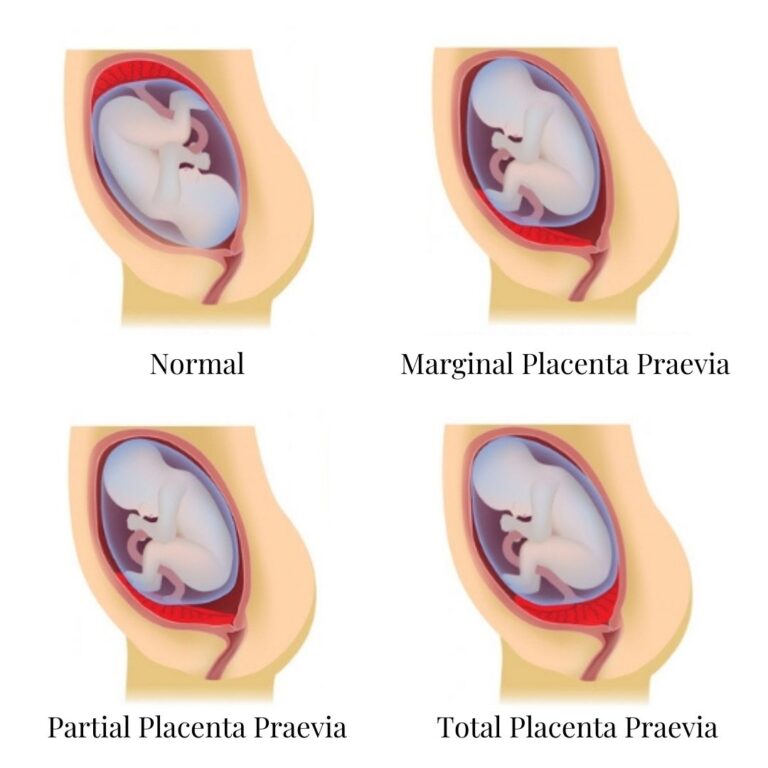
Whether the placenta is anterior, posterior, or fundal. All three are normal placental positions. Risk includes when the placenta lies near to the os which is termed as low lying placenta or placenta previa if the placenta covers the internal os.
No need to get shattered with the results. It’s important to remember that the test is not 100% accurate and it’s not a confirmed diagnosis. The NT Scan and double marker are the screening test and not diagnostic ones. These tests only predict if your baby is at high risk or low risk of having a genetic condition. Rather, let’s see an advantage of the test being taken early in the pregnancy so that it gives you time to carefully consider your next steps and get the support and advice you need.
Further, your doctor will counsel you regarding your options. This will include the offer of confirmatory tests ( diagnostic invasive testing) to confirm the presence of a chromosomal abnormality in the baby: either a chorionic villus sampling done between the 11th -14th week of pregnancy or amniocentesis which is usually carried out after the 15th week.
As an alternative, non-invasive prenatal testing (NIPT) is just becoming available in many of the places which are again non-invasive but more accurate than the double marker. It’s important to note that NIPT is also a screening test and not the diagnostic one.
An NT Scan is a non-invasive, safe procedure for both mother and the baby. Each and every mother should go for this scan not only to understand the risk of chromosomal abnormalities but also to know the other details of the baby’s growth. This scan will definitely give you peace of mind as a mother.
You might want to check our blog on ‘DOUBLE MARKER TEST – A LOOK INTO YOUR CHILD’S CHROMOSOMES’ which explains everything about Double marker test and down syndrome.
For many out there Diwali is the festival of lights. But If you ask me what is Diwali, I would say “Diwali is a wisely

As the best parent, everyone tries to protect their child in all possible ways. Our parents did the same to us and still do it.

Besides being a Doctor, like every other pregnant woman I too had a lot of confusion regarding what essentials I should have to make my
As a mother, I always had a hundred thoughts in my mind during my pregnancy. I always wanted to be sure about whether my baby

Are you dealing with stress in pregnancy? Have you ever desired for a quick and easiest remedy to charge you up in those depressed and

At their core, traditional recipes are a way of ensuring you’re eating what your body needs to stay healthy and feeling good. Each recipe has
Copyright © 2021 truptwellness | Powered by truptwellness
Any questions? Click for Consultation.
One Response
An NT Scan is a non-invasive, safe procedure for both mother and the baby.